

Understanding Myofascial Trigger Points: The Clinical Science Behind “Muscle Knots”
Quick answer: Myofascial trigger points (“muscle knots”) are not permanent structural lumps, but rather microscopic clusters of muscle fibers locked in a continuous contraction—a state driven by excessive acetylcholine release that pinches local capillaries, restricts blood flow, and traps inflammatory chemicals. Confirmed by NIH microdialysis and ultrasound elastography as dark, measurably stiffer nodules with reduced circulation, trigger points respond to therapies like foam rolling, targeted compression, and dry needling not by physically “breaking up” tissue, but by stimulating mechanoreceptors to signal the central nervous system to reduce muscle tone and restore healthy fluid flow.
Nearly everyone has experienced it: a persistent, dull ache in the calf, shoulder, or thigh that leads to a tender, hardened spot beneath the skin. Commonly called a “muscle knot,” its clinical name is a myofascial trigger point (MTP).
While long recognized in manual therapy, myofascial pain syndrome (MPS) and trigger points were historically met with skepticism in formal medicine due to a lack of objective imaging. Today, advanced ultrasound elastography, biomarker assays, and electrophysiological studies provide clear evidence of what trigger points are, why they form, and how to treat them effectively.

1. What Is a Myofascial Trigger Point?
A myofascial trigger point is a hyperirritable spot within a taut band of skeletal muscle fibers. Clinically, trigger points are categorized into two primary types:
Active Trigger Points: Cause spontaneous, continuous pain both at rest and during movement. They produce a characteristic pattern of referred pain—discomfort felt in an area distant from the actual knot.
Latent Trigger Points: Painless during ordinary movement, but tender when compressed. Although silent, they can restrict range of motion, alter muscle activation patterns, and cause localized muscle weakness.
The Integrated Hypothesis
The prevailing pathophysiological explanation for trigger points is the Integrated Hypothesis, first proposed by Drs. David Simons and Janet Travell and expanded by Dr. Robert Gerwin.
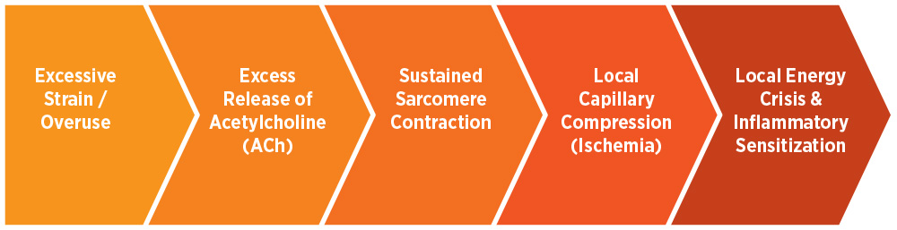
Excessive Strain / Overuse: Physical overload—whether from acute trauma, repetitive micro-injury, or sustained postural strain—places unaccustomed mechanical stress on muscle fibers. This overload damages cell membranes and triggers dysfunctional signaling at the neuromuscular junction, serving as the primary catalyst for the entire cascade.
Abnormal Motor Endplate Activity: Acetylcholine (ACh)—the primary chemical messenger that allows the nervous system to signal muscle movement—leaks excessively at the neuromuscular junction due to tissue strain. Under normal conditions, ACh triggers brief, controlled contractions before being rapidly broken down; however, this excessive release causes persistent depolarization of the muscle cell membrane, keeping the muscle fiber continuously activated.
Sustained Sarcomere Contraction: Calcium remains trapped inside the muscle cell, causing adjacent sarcomeres (the microscopic contractile units of muscle) to lock in a shortened state.
The “Energy Crisis”: This continuous contraction compresses local capillaries, severely restricting blood flow (ischemia) and oxygen delivery (hypoxia).
Sensitization: Deprived of oxygen and nutrient exchange, the tissue enters a localized energy crisis. The area accumulates inflammatory biochemicals—such as substance P, calcitonin gene-related peptide (CGRP), and bradykinin—which lower the threshold of local nociceptors (pain receptors), creating a highly sensitized, painful spot.
2. How Trigger Points Are Visualized and Diagnosed
For decades, diagnosis relied entirely on physical palpation—identifying a taut band, finding a tender nodule, and eliciting a local twitch response (LTR), a brief involuntary contraction of the muscle fibers.
Modern diagnostic technology has confirmed these physical findings:
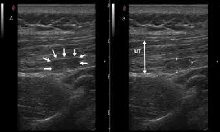
Diagnostic Ultrasound & Sonoelastography: Studies published in journals such as the Archives of Physical Medicine and Rehabilitation show that active trigger points appear on high-frequency ultrasound as distinct, hypoechoic (darker) nodules. Elastography demonstrates that these spots are significantly stiffer and less elastic than surrounding healthy muscle tissue.
Microdialysis Biomarker Profiling: Research led by the National Institutes of Health (NIH) used microdialysis needles to sample the biochemical environment of active trigger points. The samples revealed significantly elevated concentrations of inflammatory mediators, neuropeptides, and a lower (more acidic) pH compared to non-affected muscle tissue.
Doppler Imaging: Vascular Doppler studies confirm decreased arterial blood flow (increased vascular resistance) directly inside the trigger point during contraction, validating the ischemic nature of the local energy crisis.
3. Proven Treatment Strategies: What the Research Shows
Effective treatment of myofascial trigger points requires disrupting the sustained contraction cycle, restoring local blood flow, and retraining muscle mechanics.
| Treatment Modality | Primary Mechanism | Evidence Level |
| Self-Myofascial Release (SMR) | Neurosensory modulation; reduces motor neuron excitability and temporary improvement in vascular perfusion. | Moderate-High (effective for short-term relief and mobility) |
| Dry Needling | Mechanical disruption of dysfunctional endplates; elicits a local twitch response that depletes inflammatory chemicals. | High (supported by multiple systematic reviews for acute pain) |
| Targeted Ischemic Compression | Sustained manual pressure deprives tissue temporarily, triggering a rush of fresh blood (hyperemia) upon release. | Moderate-High |
| Acoustic Wave / Shockwave Therapy | Extracorporeal shockwaves stimulate microcirculation, reduce substance P, and promote tissue remodeling. | Moderate (growing clinical adoption) |
Mechanisms of Self-Care (Foam Rolling & Massagers)
Contrary to popular belief, self-myofascial release (SMR) with foam rollers, massage sticks, or therapy balls does not physically “break up” scar tissue or physically lengthen dense fascia. High-resolution biomechanical studies demonstrate that human fascia requires thousands of pounds of force to structurally deform.
Instead, SMR works primarily through neurological and vascular pathways:
Central Nervous System Modulation: Deep, sustained pressure stimulates mechanoreceptors (Ruffini and Pacinian corpuscles), which send inhibitory signals to the central nervous system, down-regulating sympathetic tone and relaxing the muscle.
Transient Rehydration: Compression squeezes fluid out of congested fascial layers; when released, fresh blood and interstitial fluid flood back into the tissue, improving fascial glide and nutrient exchange.

4. Key Factors in Preventing Recurrence
Treating a trigger point offers temporary relief if the underlying drivers remain unaddressed. Research indicates that the primary drivers of chronic myofascial pain include:
Postural Strain & Ergonomics: Sustained, low-level static contractions (e.g., sitting at a desk with elevated shoulders or poor head alignment) keep motor endplates continuously active without adequate rest windows.
Repetitive Overload: Unaccustomed exercise intensity or repetitive strain tasks create micro-trauma across sarcomeres faster than they can repair.
Dehydration & Electrolyte Imbalance: Inadequate hydration impairs fascial fluid dynamics, making tissues more susceptible to friction and tightness.
Sleep Deprivation & Stress: Elevated cortisol levels and sympathetic nervous system activation lower systemic pain thresholds and increase resting muscle tone.
Clinical Summary
Myofascial trigger points are not imaginary, nor are they permanent structural knots. They represent a well-documented physiological state of localized sarcomere shortening, local ischemia, and neuro-chemical sensitization.
Whether addressed through professional interventions like dry needling and physical therapy, or self-management techniques like sustained compression, gentle stretching, and ergonomic adjustment, the goal remains the same: restore blood circulation, reset neuromuscular tone, and allow the tissue to return to a healthy, elastic state.
Frequently Asked Questions
What actually is a "muscle knot"?
A “muscle knot” is the common term for a myofascial trigger point (MTP). It is a hyperirritable spot within a tight, rope-like band of muscle tissue where microscopic contractile units (sarcomeres) become locked in a continuous state of contraction.
What is the difference between an active and a latent trigger point?
Active Trigger Points: Cause constant, spontaneous pain even when you are resting. They frequently produce referred pain—discomfort felt in a completely different area of the body than where the knot is located.
Latent Trigger Points: Only hurt when direct pressure is applied to them. While they don’t hurt continuously, they can still cause muscle stiffness, weakness, and restricted movement.
What causes a trigger point to form in the first place?
Mechanical Strain: Muscle overuse, repetitive micro-trauma, or poor posture overloads the tissue.
Acetylcholine Surge: The body releases excess acetylcholine (ACh) at the neuromuscular junction.
Sarcomere Lock: Sarcomeres continuously contract and trap calcium inside the cells.
Ischemia (Reduced Blood Flow): The persistent contraction squeezes nearby capillaries, cutting off local blood and oxygen supply.
Energy Crisis & Pain: Lacking oxygen, the tissue enters a metabolic crisis. Inflammatory chemicals (like substance P and bradykinin) pool in the area, lowering the activation threshold for pain receptors.
Can you see a muscle knot?
Rarely with the naked eye, but clearly with medical imaging.
To the naked eye, a muscle knot rarely looks like a distinct bump or ball under the skin. Instead, you might only see subtle secondary signs, such as a localized twitch when the spot is pressed, minor asymmetry in a muscle belly, or temporary surface redness after massage.
However, advanced diagnostic imaging easily visualizes what is happening beneath the surface:
Diagnostic Ultrasound & Elastography: Reveals active trigger points as distinct, darker (hypoechoic) nodules that are measurably stiffer and less elastic than surrounding healthy muscle.
Doppler Imaging: Shows decreased arterial blood flow directly inside the contracted knot due to microvascular compression.
Microdialysis: Samples taken directly from active knots confirm elevated levels of inflammatory neuropeptides and a lower (more acidic) tissue pH.
Does foam rolling physically "break up" scar tissue or muscle knots?
No. Human fascia and muscle tissue require thousands of pounds of force to structurally deform or “break apart.” Instead, self-myofascial release (SMR) tools work primarily through neurological and vascular mechanisms:
Nervous System Reset: Sustained pressure stimulates mechanoreceptors that send signals to the central nervous system to down-regulate muscle tone and relax the area.
Fluid Exchange: SMR acts like squeezing a sponge—releasing the pressure allows fresh, oxygenated blood and fluid to rush back into congested tissue.
What are the most effective treatments for trigger points?
Evidence-based approaches focus on restoring blood flow and resetting nerve signals:
Self-Myofascial Release (SMR): Sustained compression using foam rollers, therapy balls, or massage tools for temporary relief and improved mobility.
Dry Needling: Insertion of fine needles directly into the trigger point to elicit a local twitch response (LTR), which resets dysfunctional endplates and flushes out inflammatory chemicals.
Ischemic Compression: Manual pressure held over the spot to temporarily interrupt blood flow, triggering a surge of fresh circulation upon release.
Addressing Underlying Factors: Adjusting posture, improving ergonomics, staying hydrated, and correcting movement mechanics to stop recurring strain.
Medical disclaimer: This article is for general informational and educational purposes only and is not a substitute for professional medical, veterinary, or fitness advice, diagnosis, or treatment. Always consult a qualified professional before starting a new exercise, recovery, or treatment program, or if you have any concerns about a health condition.
Safety disclaimer: Always read and follow the manufacturer’s instructions, warnings, and usage guidelines for any product or equipment mentioned here. Use tools and techniques as directed and within your own ability; start gently, stop if you feel pain or discomfort, and seek professional guidance if you are unsure. Improper or excessive use can result in injury.